|
|
Cortical Desmoid
Distal Femoral Metaphyseal Irregularity, Avulsive Cortical Irregularity, Periosteal Desmoid
General Considerations
- AKA Distal Femoral Metaphyseal Irregularity, Avulsive Cortical Irregularity, Periosteal Desmoid
- Age range: 3-17 years, most common 10-15
- Cortical irregularity at the posterior, medial and distal femur deep to the attachments of medial gastrocnemius or distal adductor magnus
- Incidental lesion
- May move away from epiphysis with age and growth
- Most likely due to chronic avulsions
Clinical Findings
- Incidental finding not to be confused with something more serious
Imaging Findings
- Cortical irregularity
- Occurs in a classical location, posterior, distal femur
best seen on lateral view of knee
- May present as an oval lucency on frontal view
- Concave and sclerotic border
- No soft tissue mass
- Bilateral 25%; more often on left
- May have solid periosteal reaction adjacent to lesion
- May contain calcifications on CT
- MR: Intermediate T1/heterogeneous T2; may enhance on T1
Differential Diagnosis
- Osteosarcoma
- Osteomyelitis
- Metastases
Treatment
- Don’t touch--No treatment needed
Prognosis
- Disappears after epiphyses close

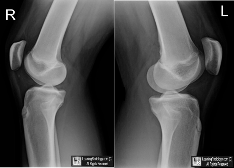
Cortical Desmoid. Note the cortical irregularity of the distal, posterior femur on the left side (yellow arrow) with no associated bone destruction or soft tissue mass. Although frequently bilateral, the right knee in this patient is normal (white arrow) and demonstrates the normal appearance of the cortex.
For this same photo with the arrows, click here
For more information, click on the link if you see this icon 
|
|
|
